
A group of scientists from Wrocław University of Science and Technology, led by Dr. Habil. Krzysztof Opieliński, has developed an innovative, non-invasive and harmless method for diagnosing breast cancer. Their collaboration with the Olsztyn-based company Dramiński S.A. has resulted in the creation of an innovative ultrasound tomograph that combines the diagnostic capabilities of mammography, ultrasound and magnetic resonance imaging.
The device will enable fast and inexpensive screening for breast cancer. Bolesławiec resident Mateusz Celmer is among its designers.
“At the heart of our device is a ring-shaped ultrasound array consisting of 1,024 miniature piezoceramic transducers, enabling the generation and reception of ultrasound vibration pulses in appropriate sequences. It allows the entire breast, immersed in water, to be scanned from multiple directions and at different levels, followed by processing the data obtained and reconstructing two- or three-dimensional ultrasound images of any breast cross-sections,” explains Dr. Eng. Mateusz Celmer, a member of the Wrocław University of Science and Technology team. “An examination performed with such a hybrid ultrasound tomograph makes it possible to assess the risk of malignancy. The examination is fast (about 10 minutes), non-invasive, safe and inexpensive. Patients are not exposed to harmful radiation at all, because our device uses only harmless ultrasound waves, like those used in ultrasound examinations.
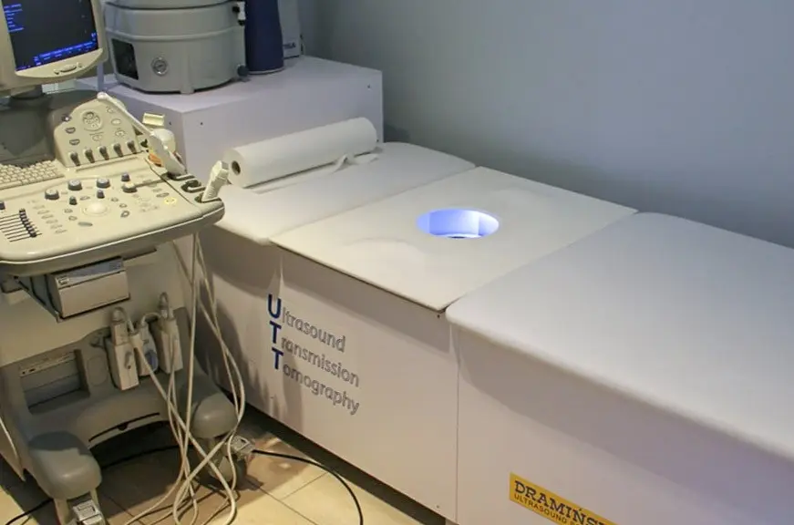
The work is funded by the Olsztyn-based company Dramiński, partly במסגרת an implementation grant from the National Centre for Research and Development, financed by the European Regional Development Fund under the “Smart Growth” programme. According to the plans, the device should enter the market at the end of 2018.
Together with Dr. Habil. Krzysztof Opieliński, professor at Wrocław University of Science and Technology, the tomograph was developed by Prof. Habil. Eng. Tadeusz Gudra, MSc Eng. Juliusz Bednarek, Dr. Eng. Piotr Pruchnicki, Dr. Eng. Mateusz Celmer and MSc Eng. Tomasz Świetlik.
Breast cancer is the most common cancer among women in Poland and ranks second in terms of mortality. However, the disease is curable when detected early. The effectiveness of early breast cancer detection using ultrasound is estimated at 50%, while that of standard mammography (MMG) is approximately 80%. Nevertheless, this method involves compressing the breast, which can be painful, as well as harmful exposure to X-rays. According to the American Cancer Society, examinations using this method carry a sufficiently low risk of error only in women over 45, when breast tissue is less dense and contains more fat, minimizing the risk of missing a lump. The most effective method currently available is magnetic resonance imaging (MRI), which can detect even 2-millimetre cancerous lesions. The main disadvantages of this method are the enormous cost of purchasing and maintaining an MRI scanner, as well as the need to administer a contrast agent intravenously, which can cause allergic reactions and is harmful to people with kidney and liver diseases. Therefore, there is no widely available, reliable screening method.